Detailed Images Help Identify Cancer Stage
Colorectal cancer is the third most common cancer in both men and women in the United States, with more than 100,000 new cases diagnosed annually. It also has a high recurrence rate, with 30% to 40% of patients developing new cancers within a five-year period.
To diagnose recurring cancers as early as possible, patients are advised to have regular imaging tests, including colonoscopies. When signs of recurrence to occur, more sensitive tests are required. At Main Street Radiology, we often use positron emission tomography (PET) scans to help diagnose colon cancer and identify the stage to which it has progressed.
With PET scans, patients ingest or receive injections of radioactive dyes, which collect in parts of the body with diseased tissue and help doctors collect clear images of the affected area. Studies show that this technology can be more effective than CT scans or MRIs in determining the extent of disease.
Case History: Status-post sigmoid colon resection and increasing CEA level. CT examination was performed (Figure 1) demonstrating soft-tissue structure adjacent to the colon anastomosis (arrow), which may represent recurrent tumor or fibrosis. The patient was referred to Main Street Radiology for a PET scan.
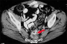
Figure 1
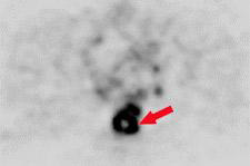
Figure 2
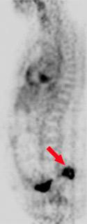
Figure 3
Findings: Axial (Figure 2) and sagittal (Figure 3) PET images demonstrate hypermetabolic activity (arrows) corresponding to the CT finding, compatible with local recurrent tumor. There was no evidence of metastatic disease elsewhere.
Discussion: PET scanning has been widely established as the most accurate non-invasive test for staging and re-staging colorectal carcinoma. For hepatic metastasis, PET is superior to CT, with sensitivity of 88% vs. 38% and specificity of 100% vs. 97% (Radiology 1998; 206:755-760). PET is also highly accurate at demonstrating recurrent colorectal cancer in patients who have indeterminate findings at CT or MR, with sensitivity and specificity for local pelvic recurrence at 95% and 97% respectively (J Nucl Med 2000; 41:1177-1189). PET allows differentiation of recurrence from fibrosis, superior to CT, with sensitivity of 93% vs. 60%, and specificity of 97% vs. 72 % (Eur J Surg Oncol 1995; 21:517-522).
PET has been approved for Medicare reimbursement for the diagnosis, staging, and restaging of colorectal cancer. During the initial staging of disease, PET plays a valuable role in determining the true extent of disease, and to help plan surgical and non-surgical therapeutic procedures. PET is also valuable in re-evaluating patients following treatment, with or without rising CEA level.
PET offers information not available through other type of diagnostic studies. Since PET tracer FDG is taken up at a cellular level, functional images are generated that complement the traditional anatomic images generated through CT and MRI studies.