Additional Testing Can Result in More Accurate Diagnosis
One out of every eight American women get breast cancer sometime during their lives. Of those who survive the initial encounter, a significant number are diagnosed with the disease again in the future, with the exact percentage depending on the precise previous diagnosis and treatment.
Accurately diagnosing breast cancer - and determining its stage – in patients who have already had it brings its own set of challenges. Traditional CT scans often have trouble distinguishing between new cancers and anatomical changes brought about by the first battle with the disease. At Main Street Radiology, we often use positron emission tomography (PET), which involves the injection of a radioactive dye to produce high-quality images, to assist in the diagnostic process.
History: A 62-year-old female who had breast cancer surgery and subsequent radiotherapy two years before was referred to Main Street Radiology for tumor re-staging.
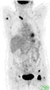
Figure 1
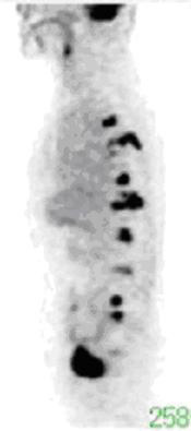
Figure 2
Findings: Coronal (Figure 1) and sagittal (Figure 2) whole body images demonstrate multiple foci of increased activity (black) in the spine, right axilla, and both inguinal regions, compatible with metastatic disease. Normal physiological activity is seen in the brain, heart, and urinary bladder.
Discussion: Accurate staging of breast cancer recurrence is critical for therapeutic planning. Traditional non-invasive imaging include CT and bone scan. Limitations of CT include the difficulty in distinguishing post-operative and post-radiation changes from recurrence, and inability to identify and characterize small lesions. In addition, detection of lesions is limited to the anatomic region being imaged, as a whole-body CT is not typically performed. Bone scan, although a sensitive study for skeletal metastases, results frequently in false positive diagnoses.
Whole-body positron emission tomography (PET) has been shown to be both more sensitive and specific than CT for the diagnosis of recurrent breast cancer. It has also been shown to be more specific than bone scan. Although PET is more sensitive than bone scan for lytic bone metastases, it is less sensitive for the less common osteoblasic lesions. Therefore, bone scan should still be performed as complementary study to the PET scan. (Radiographics. 2002;22:5-17)
In the initial diagnosis of breast cancer, PET scan should not replace breast biopsy, when warranted by mammographic, ultrasound, MRI, or clinical findings. In addition, PET is less sensitive than surgical axillary node staging. PET is reserved for patients with clinical suspicion of metastatic disease or local recurrence, where PET has been shown to be superior to CT.