Radioactive Dye Helps Provide Clearer Images
Lymphoma is cancer that begins in the white blood cells of the lymph nodes, spleen, thymus, bone marrow, and other body parts. One common form is Hodgkin's Lymphoma, which accounts for about 4% of all U.S. cancer diagnoses. It is also one of the most common cancers among children, teenagers and young adults.
Thanks to progress in treatment, the survival rate for patients diagnosed with early stage lymphoma is about 85%. But like all cancers, accurate initial staging and effective tracking of a patient's response to treatment are critical.
While CT and MRI scans have often been used, Main Street Radiology has found that full-body positron emission tomography (PET) scans be a better option. With PET scans, patients ingest or receive injections of radioactive dyes, which help doctors collect clear, detailed images of diseased areas.
Case History: A 23-year-old male was diagnosed with a large mediastinal mass seen through an outside chest X-ray (Figure 1) and CT (Figure 2). A biopsy indicated Hodgkin's Lymphoma. The patient was referred to Main Street Radiology for whole body PET scan.
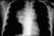
Figure 1
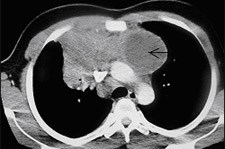
Figure 2
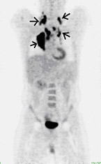
Figure 3
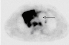
Figure 4
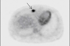
Figure 5
Findings : Coronal whole body image (Figure 3) demonstrates multiple hypermetabolic areas (dark) in the mediastimum and neck (small arrows), compatible with lymphadenopathy. Normal physiological activity within the urinary bladder is seen.
On the axial image of the chest (Figure 4), extensive mediastinal lymphadenopathy is seen corresponding to the CT finding. A photopenic (light) area is seen in the mediastinum (arrow on Figure 4) corresponding to necrosis seen on CT (arrow on Figure 2). Abnormal right internal mammary lymph node is also seen on PET (Figure 5), which was not appreciated on CT.
Discussion: Numerous clinical studies have shown that PET is the best imaging modality for lymphoma, superior to CT, MR, and gallium scan. In the management of lymphoma, PET is indicated for initial staging as well as to monitor response to treatment and to exclude recurrence. PET is especially useful in distinguishing tumor from necrosis and fibrosis.