Fusion Combines Best Features of Two Types of Scans
Often, the best diagnostic result comes from using more than one type of radiology technology. For example, one highly accurate way to identify growths on the lungs is to electronically combine - or fuse - images from a positron emissions tomography (PET)) scan with those of a CT scan.
The resulting detailed images help doctors pinpoint the location of the cancerous lung nodules and identify the proper course of treatment. At Main Street Radiology, we have acquired CT-PET fusion software. When a CT scan is taken, the data can be fused with the corresponding PET scan for the best results.
Case History: A CT examination of the chest (Figure 1a) on an 86-year-old male showed a right lung nodule. The patient was referred to Main Street Radiology for a whole body PET scan.
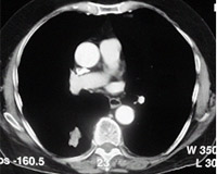
Figures 1a
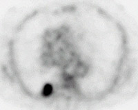
Figure 1b
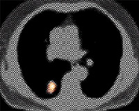
Figure 1c
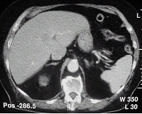
Figure 2a
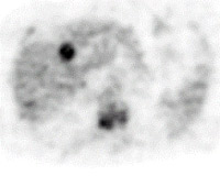
Figure 2b
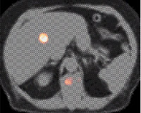
Figure 2c
Findings: A hypermetabolic focus is seen within the right lower lobe on the axial PET image of the chest (Figure 1b) corresponding to the CT finding, compatible with carcinoma. CT-PET fusion image (Figure 1c) shows CT anatomical correlation of the functional PET finding. Axial PET image of the upper abdomen (Figure 2b) along with the CT-PET fusion image (Figure 2c) show abnormal activity within the liver and spine (arrows), compatible with metastatic disease, which are not appreciated on the CT exam (Figure 2a).
Discussion: PET is a functional imaging study, which exploits the hypermetabolic activity of cancers, manifested by increase uptake of a radioactive glucose analogue. However, since most normal tissues demonstrate far less activity, anatomical detail is limited. Correlation with a CT scan is often helpful to localize the exact location of PET scan abnormalities. While side-to-side comparison of PET and CT scan is usually sufficient, "fusion" of the two studies into a single set of images can be helpful.