Deficits in Certain Brain Activity Revealed by Testing
Almost everyone knows about the devastating affects of Alzheimer's Disease on individuals and families. While there is no cure for this form of dementia, there are now drugs that can slow its progression. That makes early detection critically important.
Traditional CT and MRI scans each have drawbacks in diagnosing Alzheimer's. At Main Street Radiology, we use sophisticated positron emission tomography (PET) scans, which utilize radioactive tracers to identify metabolic deficits and protein plaque in the brain that are indicators of the disease. By identifying these signs, doctors can diagnose Alzheimer's earlier, even before symptoms appear.
History: Doctors found signs of early onset-Alzheimer's disease in a 34-year-old male.
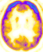
Figure 1
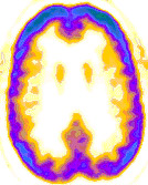
Figure 2
Findings: Axial PET image of the brain (Figure 1) demonstrates normal activity in the frontal lobes (bright blue), with diminished activity in the parietal, temporal, and occipital lobes. This pattern is typical of Alzheimer disease. As a comparison, Figure 2 is of a different patient with normal brain activity.
Discussion: Conventional imaging with CT and MR is neither sensitive nor specific for the diagnosis of Alzheimer disease (AD). The role of CT and MR in the work-up of dementia is primarily to assess the degree of atrophy and to exclude other causes of dementia, such as normal-pressure hydrocephalus, multi-infarct dementia, and intracranial mass.
PET has been used in scanning the brain by evaluating the metabolic uptake of glucose-analogue FDG. High accuracy in differentiating Alzheimer's Disease from other forms of dementia has been reported. In patients with Alzheimer's, deficits in temporoparietal metabolism are typically seen. Sensitivity of 94% has been reported in the literature (J Nucl Med 1994; 35:391-398), with high accuracy seen in early and mild forms of the disease. Abnormal temporoparietal uptake has also been demonstrated in asymptomatic patients with family history of early-onset AD (Ann Neurol 1997; 42:85-94). In a multinational consortium study (JAMA 2001; 286:2120-2127), PET accurately predicted cognitive decline in patients with Alzheimer's, with sensitivity of 93%.
In 2004, Medicare approved coverage for PET scans in the early diagnosis of Alzheimer's Disease. This is an important step, as new drugs work best in the early stages to slow the progression of this disease.
PET offers information not available through other types of diagnostic studies. Since PET radioactive tracer FDG is taken up at a cellular level, functional images are generated that complement the traditional anatomic images generated through CT and MRI studies.